Retinal Surgery
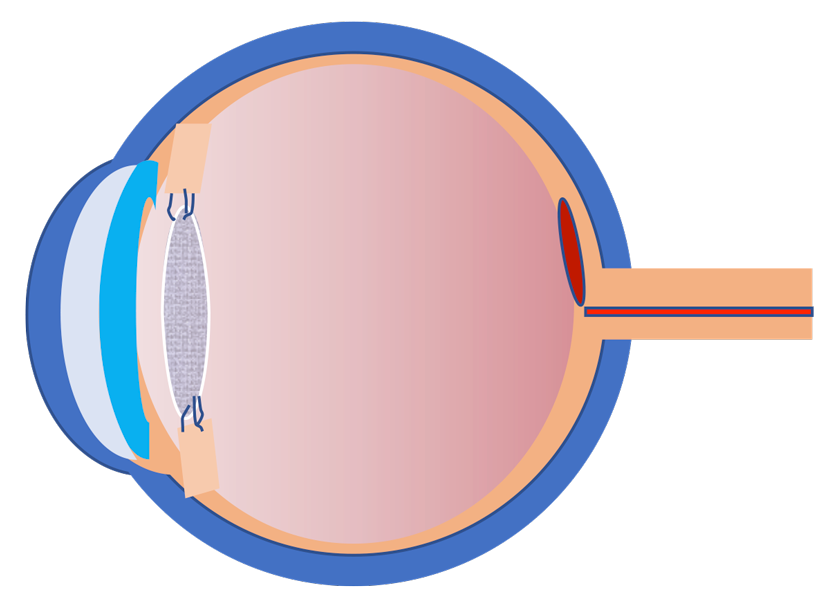
Eye structure
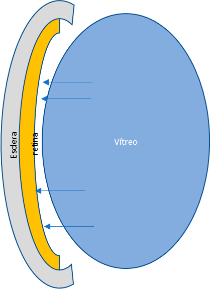
The retina, therefore, is a thin layer strongly adhered to the eye inside.
But what keeps it stuck?
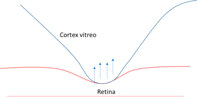
Normally the vitreous cortex, which constitutes the outer part of the vitreous, is firmly adhered to the inner walls of the eye, and therefore, in direct contact with the retina.
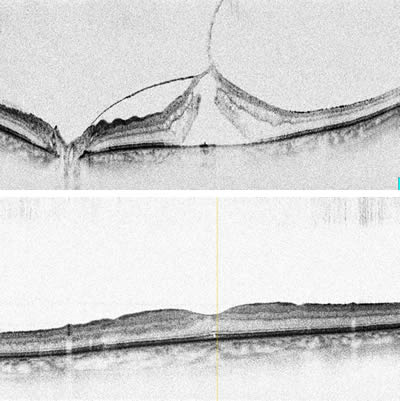
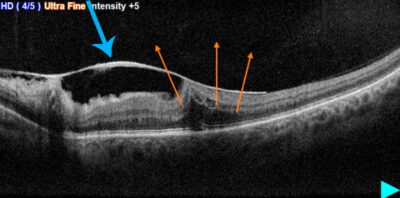
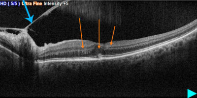
Full Macular Hole
resolution.
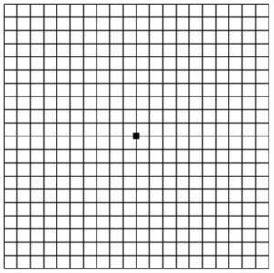
This is probably the least desired situation. The patient then perceives a black spot in the center of his visual field and loss of acuity that is variable but always severe.
This pathology can also be treated surgically by releasing traction in the area surrounding the hole. In most cases the hole is closed with this procedure.
But it is convenient for the patient to know that the closure of the hole does not mean the restoration of vision, although in most cases it does occur over the months following the treatment.
Epiretinal Membrane
Its frequency is approximately 2% in people over 50 and increases with age. In most cases the cause is unknown. In this case, the difference with the Vitreous Traction that we have talked about before is that the detachment has been complete, however a dense membrane is formed on the surface of the retina, which when contracted deforms the retina and therefore there is no upward traction, as in the tractional syndrome, but tangential, that is, sideways, causing wrinkling of the retina on which it is located. The symptoms are similar to those of vitreomacular Traction syndrome.
Why surgery at Sol Eyes?
To offer you the best services, not only do we provide you with world-class eye care in our clinics, but also with a large range of top medical specialists, with the most advanced examination and surgical equipment, as well as the experience gained from decades of treating patients throughout Europe.

